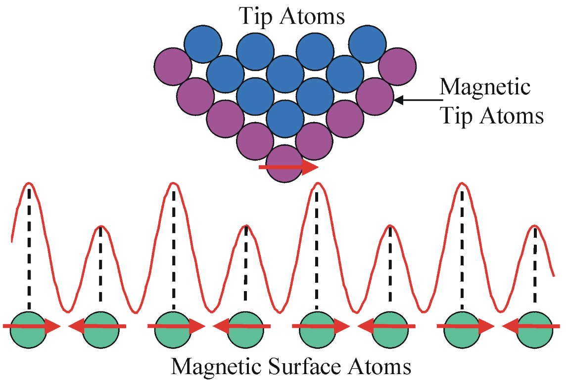
The basic idea is to explore clean and freshly-prepared thin film surfaces under ultra-high vacuum conditions which can be engineered and synthesized using molecular beam epitaxy and then investigated in-situ using scanning tunneling microscopy. Of great use in the lab is the technique of spin-polarized scanning tunneling microscopy (SP-STM) which involves using a very sharp probe tip which is also magnetic via coating a W tip with magnetic atoms such as Fe, Mn, or Cr atoms. This then makes it possible to explore the spin structure of the surfaces. I am particularly interested in using SP-STM for studying the atomic- and nanometer-scale magnetic structure of surfaces.
The lab furthermore specializes in all types of nitride systems and has previously studied thin film layers of GaN, ScN, CrN, FeN, MnN, Mn3N2, Mn4N, MnGaN, and CrGaN. Preparation of bi-layer systems such as Fe/CrN leads to being able to investigate complex magnetic properties arising from interfacial effects such as interfacial coupling.
Main research interests in the lab include:
1) quantum properties of complex materials and low-dimensional systems
2) electronic and magnetic properties in nitride systems
3) magnetic properties of transition metal and rare earth nitride systems
4) room-temperature ferromagnetism in 2-Dimensional MnGaN (MnGaN-2D)
5) spin Kondo (zero bias) resonances in ferromagnetic islands on magnetic nitride surfaces
Additional research interests:
6) Skyrmions in magnetic nitride systems
7) Novel quantum states such as Majorana zero modes in nitride materials
Phases of Research Projects
In the research performed in the group, there are often 4 stages to a successful project as shown in the following diagram. First we explore the growth of the material; second, we investigate the structural properties; once this is determined, it is possible to look into the electronic and spin magnetic properties. Magnetic properties can be closely related to the electronic properties as well.
Material Synthesis and Analysis Techniques
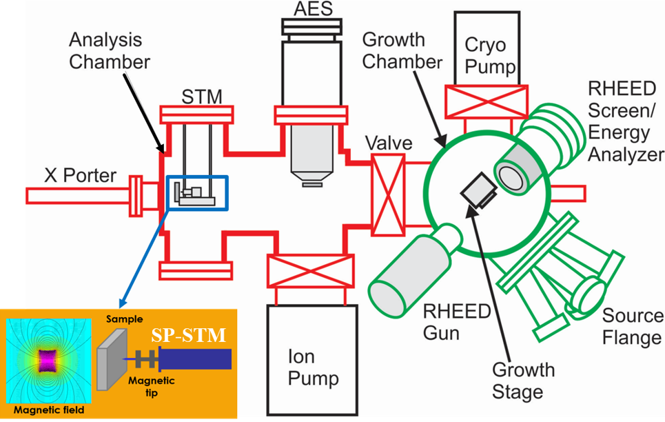
New materials are frequently synthesized using molecular beam epitaxy. MBE growth involves co-deposition of one or more elemental materials from high temperature furnaces (effusion cells) onto a crystalline substrate such as sapphire (0001) which is usually heated to a certain growth temperature T_S. The deposition rate is calibrated using a crystal thickness monitor, and the progress of the growth is monitored in real time using reflection high energy electron diffraction (RHEED). RHEED gives reciprocal-space information about the quality of the surface including its smoothness and crystallinity.
After growth is finished, the sample is transferred through vacuum into the adjoining scanning tunneling microscopy (STM) chamber and then studied by STM. STM gives detailed real-space views of the atomic-scale surface structure. Using these techniques in tandem comprises a powerful approach to understanding the relationship between surface formation and resultant surface properties.
Low-temperature Spin-Polarized Scanning Tunneling Microscopy
In order to make full use of spectroscopic techniques such as SP-STM, my group has developed a second laboratory with a dedicated low-temperature SP-STM having a variable in-situ, applied magnetic field. The facility is coupled to a custom, state-of-the-art materials synthesis facility for exploration of new material systems.
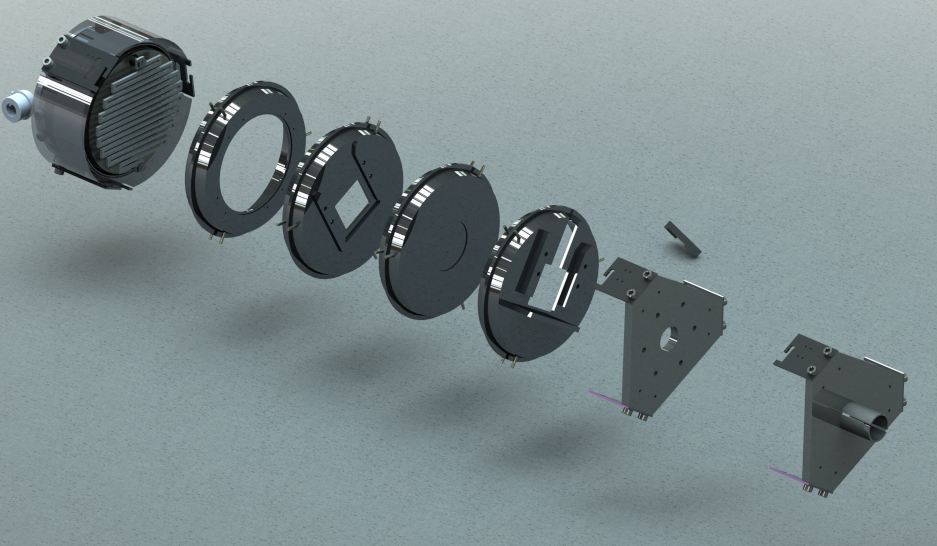
Exploding View of the Bayonet Style Sample Transfer System in Lab 102