|
 |
|
Lawrence M. Witmer,
PhD
Professor of Anatomy
Chang Professor of Paleontology
Dept. of Biomedical Sciences
Heritage
College of Osteopathic Medicine
Life Science Building, Rm 123
Ohio University
Athens, Ohio 45701 USA
Phone: 740 593 9489
Fax: 740 593 2400
Email: witmerL@ohio.edu
|
|
|
|


 |
|
|
|
| |
 |
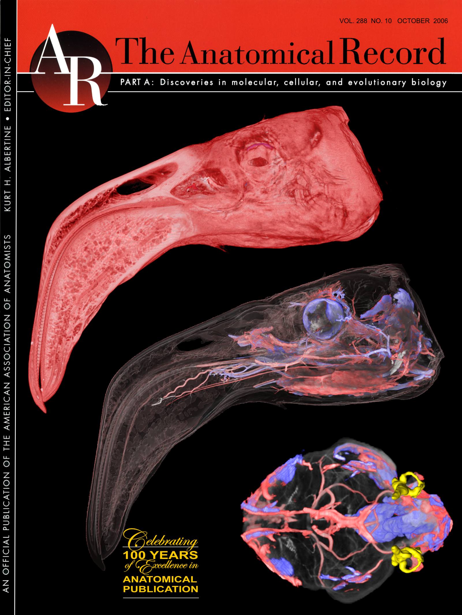
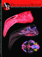
Vascular
Anatomy in Flamingos
2006. Holliday, Ridgely,
Balanoff, and Witmer. Anatomical Record |
|
|
| |
|
| |
Common Language Summary |
| |
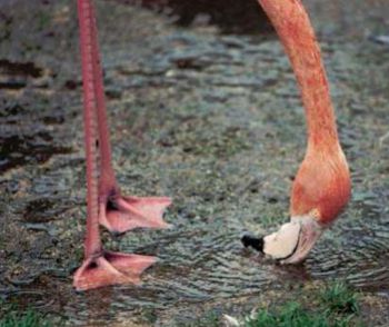
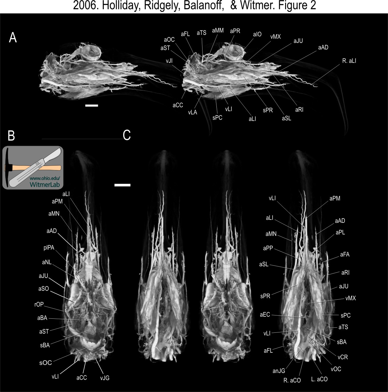
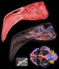
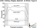
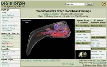
New insights into the enigmatic heads of flamingos.
Flamingos are known for their peculiar feeding behavior in which
they hold their beaks almost upside down as they filter food
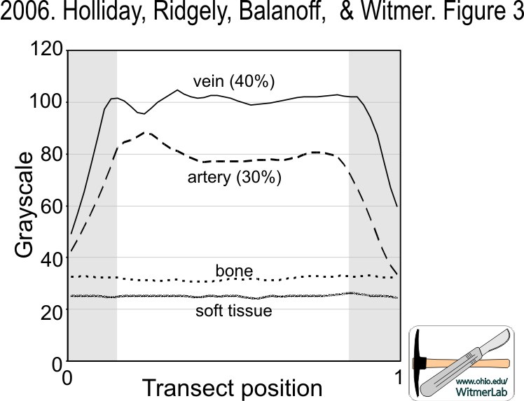
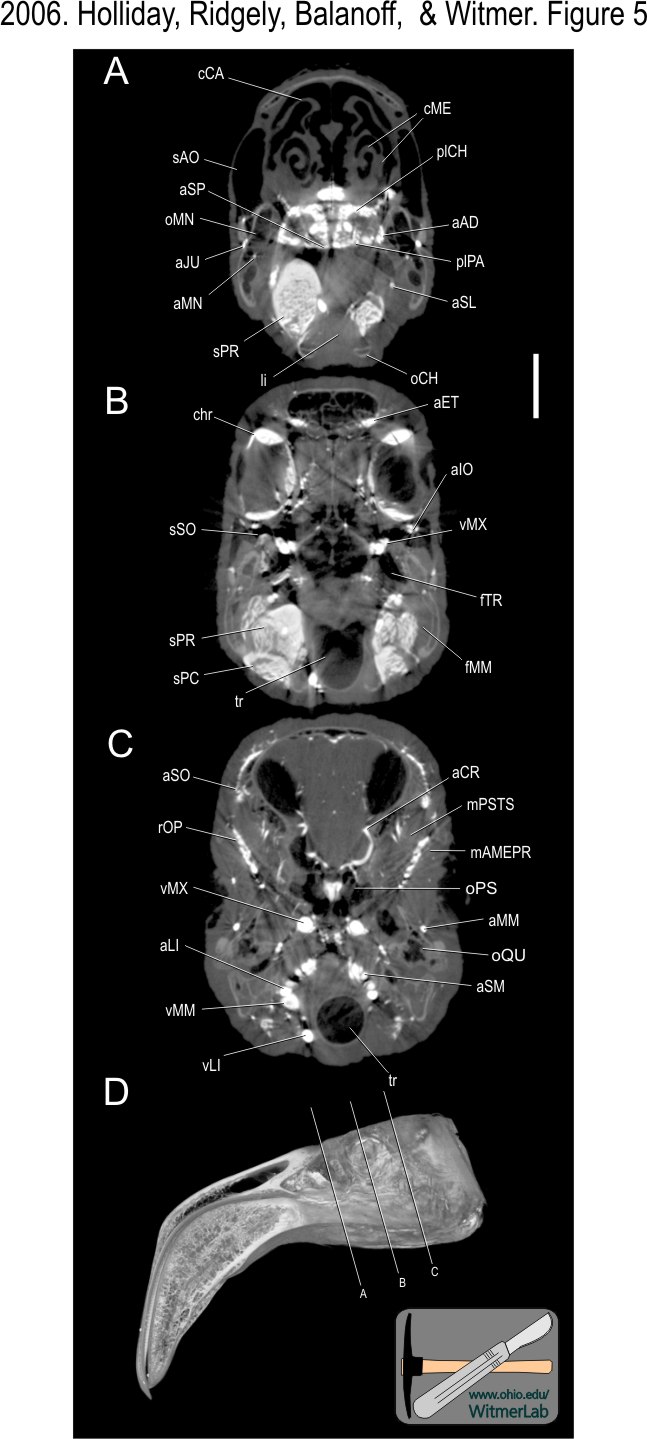
from shallow lakes. A new CT scanning technique that highlights
blood vessel anatomy allowed discovery of a large vascular
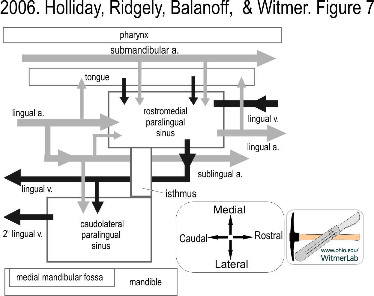
device called a “paralingual sinus” associated with the tongue
and floor of the mouth in Caribbean flamingos (Phoenicopterus
ruber). Bony evidence suggests that paralingual sinuses are
present in other species of flamingos as well but absent in
other birds, suggesting that the sinuses function in connection
with the peculiar filter-feeding style of flamingos. Flamingos
filter small food items from water using rapid, piston-like
movements of the tongue. The paralingual sinuses are composed of
erectile tissue and when engorged with blood would add
structural stiffness to the tongue and floor of the mouth,
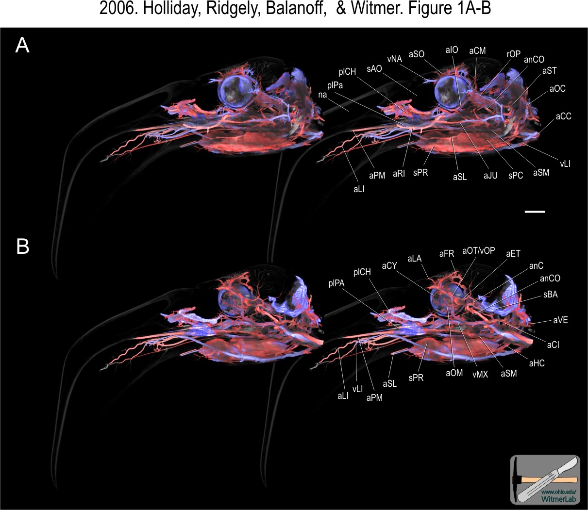
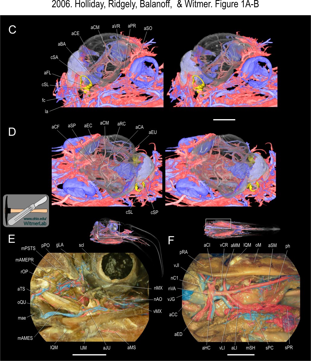
potentially improving mechanical efficiency. In addition to
these vascular devices, arteries and veins of the eye socket,
brain, and face were also illustrated using this new 3D vascular
imaging technology. |
| |
|
News Release from Ohio University |
|
Download the cover! |
Download the cover art! |
|
Complete citation and full-text download (PDF):
Holliday, Casey M., Ryan C. Ridgely, Amy M. Balanoff,
and Lawrence M. Witmer. 2006. Cephalic vascular
anatomy in flamingos (Phoenicopterus ruber) based
on novel vascular injection and computed tomographic
imaging analyses. Anatomical Record
288A(10):1031–1041. |
|

Larger versions of Figures 1A-B, 1C-F, and 2 from the original
article. |

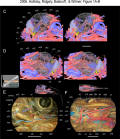
Larger versions of Figures 3, 4, and 5 from the original
article. |
|
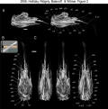
Larger versions of Figures 6 and 7 from the original
article. |
Many associated movies and images are available on
DigiMorph.org |
|

Brevard Zoo, Melbourne, Florida
Research Partner: Source of flamingo specimen |

University of Texas at Austin, HRXRCT Facility
Research Partner: CT scanning site |
| Funding for this research comes in part from the
following National Scientific Foundation (NSF) grants:
NSF IBN-0343744 to Witmer, NSF IBN-0407735 to Witmer &
Holliday, and NSF IOB-0517257 to Witmer & Ridgely. Other
funding provided by Ohio University. |
PDF of classic paper on flamingo
feeding:
1957. Jenkin. Filter-feedings in flamingos. |
|
| |
|
| |
note: Research
in the Witmer lab does not involve experimentation on live
animals. Specimens of modern animals used in research are
salvage specimens, obtained legally from commercial or
governmental sources. |
|